What are the signs of CrCL disease?
The signs of CCL rupture can be quite variable depending on the amount of damage to the ligament and meniscus but involve some degree of hind limb lameness. Hind limb lameness can appear to happen suddenly or become worse over time. Signs you may notice in your pet include a reluctance to place the limb on the ground, limping and decreased muscle mass in the affected leg. Dogs with Cranial Cruciate Ligament Disease experience pain and subsequent knee (stifle) osteoarthritis so the signs may also include a stiffness after exercise, difficulty when rising from sitting and a reluctance to jump.
Which dogs are affected?
All breeds and ages of dog are susceptible to cruciate disease. Because of the chronic, degenerative nature of CrCL disease (ageing of the ligament), middle aged to older dogs have an increased incidence of disease compared to young dogs, however mainly due to their size and conformation, some large and giant breed dogs may develop CrCL disease earlier in life.
Other factors that put your dog at higher risk for CrCL disease include:
- Obesity
- Poor physical condition
- Genetics, including poor conformation (skeletal shape and configuration)
- Concurrent disease – patella (kneecap) luxation
Because of the biomechanical nature of the disease, it will occur in both stifles of about 40-50% of affected dogs.
How is Cranial cruciate disease diagnosed?
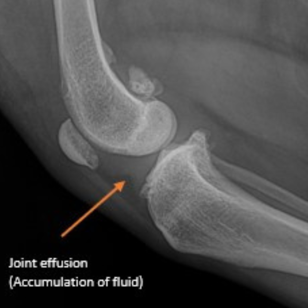
Rupture of the CrCL is diagnosed through a combination of history, a comprehensive physical examination and taking radiographs (x-rays) of the stifle. Your dog’s stifle may be swollen and is often painful to examine. These procedures will need to be performed under sedation or anaesthesia due to patient resistance and discomfort. X-rays allow your Veterinarian to:
- Confirm the presence of joint effusion (abnormal amount of fluid in the joint)
- Evaluate for the presence/degree of arthritis
- Rule out other disease conditions (like a fracture or bone cancer).

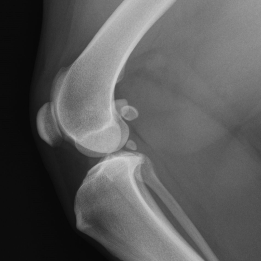
Normal Healthy Stifle

Pre-operative radiograph: cruciate ligament rupture
Definitive diagnosis of CrCL injury is made by direct visualisation of the damaged ligament during surgical investigation of the joint (arthroscopy or arthrotomy). While inspecting the joint, the surgeon will also examine all other relevant structures including the meniscus for damage and remove any damaged cartilage.
Treatment
CrCL disease is considered a surgical disease in dogs. Conservative (i.e. non-surgical) treatment is not recommended as the instability and inflammation leads to severe arthritic changes, ongoing pain and lameness. At Animalius we perform a procedure called Tibial plateau levelling osteotomy (TPLO). TPLO involves using a bone saw to make a semi-circular cut (osteotomy) in the tibia to modify the angle of the tibial plateau. The cut section is rotated and stabilised with a bone plate and screws. The procedure is designed to neutralise the biomechanical forces acting on the stifle and thus eliminates the functional need for a CrCL.
Due to the 3-dimensional nature of the stifle joint, dogs with CrCL disease may also have an increased risk of rotational instability (internal rotation) caused by a non-functioning CrCL and malalignment of the tibia and femur at the stifle joint. In severe cases, where rotational instability of the stifle joint persists following the TPLO procedure a specialised anti-rotational suture and anchor are placed, to prevent internal rotation of the stifle joint providing further stability.
Referral to Animalius and what to expect
Once your pet has been referred by your veterinarian to the team at Animalius, an appointment will be made to see our specialist surgeon. At the time of consultation our specialist surgeon will perform a comprehensive orthopaedic examination and discuss with you your pet’s surgical treatment plan.
Following consultation, the next step is for your pet to be admitted into hospital for radiographs, and surgery. The radiographs taken are to determine the extent of the disease and are used to take measurements formulating a specific surgical plan for your pet. Your Pet will then follow into surgery where an arthroscopy or arthrotomy and TPLO procedures are performed. In cases, where rotational instability of the stifle joint persists following the TPLO procedure, surgery involves the placement of a specialised anti-rotational suture and anchor.
They will stay in hospital overnight for monitoring with the plan to discharge them from the hospital the following day.
Dependant on your pet’s needs, consultation, surgical planning and surgical procedures are not always performed on the same day, this will be discussed with you at the time of booking your appointment and consultation.
Following discharge from hospital, revisit appointments are then scheduled for your pet as follows:
- Revisit consultation – 2 weeks post-surgery to check the surgical wound, assess your pet’s comfort, and remove skin stitches.
- Admit appointment – 8 weeks post-surgery. Your pet will be admitted into hospital for a physical examination, sedation and post operative radiographs. The radiographs performed determine the extent of surgical healing. Your pet will need to fast for this appointment. Results of the radiographs and a further exercise plan for your pet will be provided at the time of discharge.
Post-Operative care
Your pet will need to be kept quiet and confined for 6-8 weeks post-surgery. A small enclosure, crate or room with non-slip floors is an ideal environment. To ensure ideal conditions for healing and to encourage a successful outcome after surgery all boisterous activity including running, jumping, climbing stairs, playing with other animals, and jumping onto and off furniture must be strictly avoided during this time.
Exercise and movement are very important during the recovery period and assist in the facilitation of healing. Most patients will begin walking within 24-48 hours post-surgery and you will be provided with a tailored rehabilitation and restricted exercise program at the time of discharge. As TPLO surgery stabilises the stifle joint your pet can comfortably use the limb and exercise plans often involve supported on lead walking that encourages use of the surgery leg, ice/warm packing, massage with movement and strengthening exercises. Pain management after surgery is critical, and your pet will be required to take prescribed medications.
Post-operative complications: Post-operative complications are uncommon but can occur. Complications include bone fractures (can occur when restricted exercise and post operative care requirements are not applied correctly or are inadequate), late meniscal injury (5% for TPLO), infection (5%), rotational instability (increased risk where muscle atrophy is present, meniscectomy is required or performed, sudden onset with complete ligament rupture and knee laxity present), implant failure (rare), and soft tissue swelling around the surgery site. Some require medical management (medicine and rest). A small number of complications may require further surgery (particularly if there is deep infection, fracture, meniscal tear, rotational instability, or implant failure).
